

Retinal detachment is a serious eye emergency that can permanently affect vision if not treated quickly. The condition develops when the retina separates from the back wall of the eye, interrupting its blood and oxygen supply. Without quick treatment, retinal cells may become permanently damaged.
Recognizing the early warning signs and understanding available treatment options can help prevent severe vision loss. Modern retinal procedures, including laser therapy and surgery, have improved outcomes significantly for many patients. People searching for an experienced eye specialist in Mumbai can consult Surya Eye Hospital for timely diagnosis and retinal care.
What is Retinal Detachment?
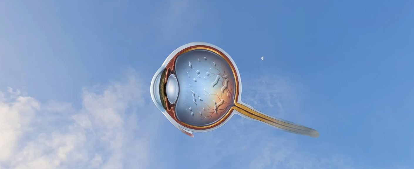
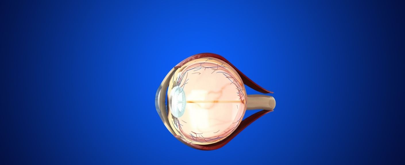
Retinal detachment is a serious eye condition in which the retina, the thin light-sensitive tissue located at the back of the eye, separates from its underlying supportive layers.
When this separation occurs, the retina cannot receive adequate oxygen and nutrients, which can rapidly affect vision and may lead to permanent vision loss if left untreated. Immediate medical attention is essential to preserve eyesight and prevent further retinal damage.
What Causes Retinal Detachment?
Retinal detachment develops due to retinal tears, eye injuries, severe myopia, diabetes, or age-related changes inside the eye. Some people may also develop retinal detachment after previous eye surgery. The common causes are explained in detail below:
Previous Eye Surgery: Patients who have undergone cataract surgery may carry a slightly increased risk of retinal detachment, especially when pre-existing retinal weakness or high myopia is present.
Age-Related Vitreous Changes: The vitreous gel inside the eye shrinks naturally with age. As the gel pulls away, it may create traction on the retina and cause tears.
Retinal Tears: A retinal tear allows fluid to enter beneath the retina. This fluid separates the retina from the underlying tissue. Understanding retinal tear vs retinal detachment is important because untreated tears can rapidly progress into full retinal detachment.
Eye Trauma: Sports injuries, falls, or accidents can directly damage retinal tissue and trigger detachment.
Diabetic Eye Disease: Advanced diabetic retinopathy may cause scar tissue formation that pulls the retina away from the eye wall.
Who Is at High Risk for Retinal Detachment?
People with high myopia, diabetes, previous eye surgery, retinal tears, or eye injuries face a greater retinal detachment risk. Aging also increases susceptibility. High-risk groups include:
- Adults above 50 years
- Patients with severe myopia
- Diabetic individuals
- Patients with a family history of retinal detachment
- Individuals with previous retinal surgery
- Patients with eye trauma history
High myopia is one of the most important risk factors because the elongated eye shape stretches and thins the retina, making it more vulnerable to tears and holes.
At Surya Eye Hospital, individuals visiting for myopia treatment in Mumbai, including LASIK surgery in Mumbai when appropriate, are advised to undergo a comprehensive retinal evaluation to detect any underlying retinal weakness before treatment.
Signs and Symptoms of Retinal Detachment
Signs and symptoms of retinal detachment include flashes of light, floaters, blurred vision, and curtain-like shadows across vision. Symptoms usually appear suddenly and may worsen quickly. Common warning signs include:
- Sudden appearance of floaters
- Flashes of light in peripheral vision
- Blurred or distorted vision
- Loss of side vision
- Shadow or curtain moving across vision
- Sudden decrease in visual clarity
An experienced eye specialist in Mumbai at Surya Eye Hospital can perform advanced retinal imaging and emergency retinal treatment for patients with acute symptoms.
Diagnosis of Retinal Detachment
Diagnosis of retinal detachment focuses on identifying retinal tears, fluid accumulation, and areas where the retina has separated from the back of the eye. Early diagnosis is important to prevent permanent vision loss. Common diagnostic methods may include:
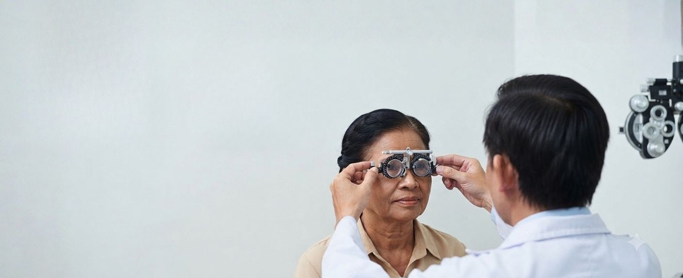
- Eye Examination: The ophthalmologist checks vision changes, eye pressure, pupil response, and symptoms such as flashes, floaters, or shadowed vision.
- Dilated Retinal Examination: Special eye drops are used to widen the pupils so the retina can be examined in detail for tears, holes, or detachment.
- Ophthalmoscopy: A specialized light and magnifying instrument helps doctors closely inspect the retina and vitreous gel.
- Optical Coherence Tomography (OCT): OCT scans provide detailed cross-sectional images of the retina to assess retinal damage and fluid buildup.
- Ultrasound Imaging: Eye ultrasound may be recommended if bleeding or cataracts block a clear retinal view.
- Visual Acuity Testing: Vision tests help evaluate the extent of vision loss or visual disturbances caused by retinal detachment.
- Medical History Review: Doctors assess previous eye injuries, surgeries, severe myopia, diabetes, or family history of retinal disorders.
Treatment Options for Retinal Detachment
Treatment options for retinal detachment include laser therapy, cryotherapy, pneumatic retinopexy, scleral buckle surgery, and vitrectomy. Doctors select treatment based on retinal damage severity and tear location.
Comparison of Retinal Detachment Treatments
| Treatment | Purpose | Best For |
| Laser Photocoagulation | Seals retinal tears | Early retinal tears |
| Cryotherapy | Freezes retinal breaks | Small retinal holes |
| Pneumatic Retinopexy | Uses a gas bubble to reposition the retina | Simple detachments |
| Scleral Buckle | Reduces retinal traction | Moderate detachments |
| Vitrectomy | Removes vitreous, pulling the retina | Complex detachments |
Retinal Detachment Laser Surgery
Retinal detachment laser surgery seals retinal tears before complete retinal separation occurs. The laser creates tiny scars around the tear to prevent fluid leakage.
Laser treatment works best during the early stages before major detachment develops.
Benefits include:
- Minimally invasive procedure
- Short recovery period
- Outpatient treatment
- Reduced progression risk
- Quick treatment time
Laser therapy may not work once complete retinal detachment develops.
Retinal Detachment Surgery
Retinal detachment surgery reattaches the retina and restores its position inside the eye. Surgeons may combine multiple procedures depending on the severity of damage.
1. Vitrectomy
Vitrectomy is a surgical procedure that removes the vitreous gel, pulling on the retina. After removing the gel, the surgeon repairs retinal tears using laser or freezing treatment. A gas bubble or silicone oil is then placed inside the eye to help keep the retina attached during healing.
2. Scleral Buckle Surgery
Scleral buckle surgery involves placing a soft silicone band around the outer wall of the eye. This band gently pushes the eye wall inward, reducing traction on the retina and helping the detached retina reattach properly. The buckle usually remains permanently inside the eye.
3. Pneumatic Retinopexy
Pneumatic retinopexy is a minimally invasive procedure used for certain retinal detachments. The doctor injects a gas bubble into the eye, which presses the detached retina back into place. Laser treatment or cryotherapy is then used to seal the retinal tear and prevent further fluid leakage.
Retinal Detachment Surgery Recovery
Retinal detachment surgery recovery may take several weeks to months, depending on the severity of retinal damage and surgical technique. Vision usually improves gradually over time.
Patients must follow postoperative instructions carefully for successful healing.
First Week After Surgery
The first week after surgery usually involves blurred vision, mild discomfort, redness, and restricted activities. Most patients require protective eye shields and medicated eye drops. Doctors may recommend rest and limited movement during early healing.
Common early recovery symptoms include:
- Mild eye pain
- Redness
- Temporary blurry vision
- Light sensitivity
- Watering eyes
Positioning Instructions
Certain retinal surgeries require strict head positioning after surgery. Proper positioning helps the gas bubble support retinal healing correctly.
Patients may need to:
- Keep the head face-down
- Avoid sleeping on specific sides
- Maintain positioning for several days
Incorrect positioning may affect surgical success.
Vision Recovery Timeline
Vision recovery after retinal surgery varies between patients. Some people improve within weeks, while others require several months.
| Recovery Period | Expected Changes |
| 1-3 Days | Redness and blurry vision |
| First Week | Mild discomfort and swelling |
| 2-6 Weeks | Gradual visual stabilization |
| 2-3 Months | Continued retinal healing |
| Several Months | Further visual improvement |
Activity Restrictions
Patients should avoid heavy physical activity during retinal healing. Excessive strain may increase eye pressure and interfere with recovery.
Doctors usually advise patients to avoid:
- Heavy lifting
- Gym workouts
- Swimming
- Eye rubbing
- Air travel is a gas bubble that remains inside the eye
Activity restrictions vary depending on the surgical technique.
Follow-Up Care
Regular follow-up visits help doctors monitor retinal healing and detect complications early. Patients should attend every scheduled appointment.
Follow-up examinations may include:
- Retinal imaging
- Vision assessment
- Eye pressure checks
- Monitoring gas bubble absorption
Success Rate of Retinal Detachment Surgery
Retinal detachment surgery has a high success rate, especially when performed early. Many patients achieve successful retinal reattachment after one procedure.
Success depends on:
- Extent of retinal damage
- Macular involvement
- Timing of surgery
- Type of detachment
- Overall retinal health
Modern surgical techniques have improved both anatomical success and visual recovery outcomes.
Factors Affecting Visual Recovery
Several factors influence postoperative vision quality.
These include:
- Duration of retinal detachment
- Severity of retinal damage
- Presence of scar tissue
- Diabetes-related retinal disease
- Patient age
Patients treated before central retinal involvement usually achieve better results.
Conclusion
Retinal detachment is a medical emergency that requires immediate diagnosis and treatment. Symptoms such as flashes, floaters, blurred vision, or shadow-like vision changes should never be ignored. Patients experiencing such symptoms should get a quick evaluation at a specialized eye hospital in Mumbai, such as Surya Eye Hospital, to prevent retinal complications.
Advanced retinal procedures, including retinal detachment laser surgery and vitrectomy, help preserve vision in many patients when performed early. Individuals with severe myopia, diabetes, or previous eye surgery should undergo regular retinal evaluations to reduce complications.
FAQs
Is retinal detachment painful?
Retinal detachment usually does not cause severe pain. Most patients notice flashes, floaters, blurred vision, or dark shadows instead. Some people may experience mild discomfort or pressure sensation. Any sudden visual change should receive immediate medical evaluation because painless symptoms can still indicate serious retinal damage.
Can retinal detachment happen suddenly?
Yes, retinal detachment can develop suddenly and progress rapidly within hours or days. Many patients first notice flashes of light, new floaters, or curtain-like shadows across vision. Immediate treatment significantly improves outcomes. Delaying medical care increases the risk of permanent retinal damage and long-term vision loss.
How long does retinal detachment surgery take?
Retinal detachment surgery usually takes one to three hours, depending on surgical complexity. Procedure duration varies according to retinal tear size, detachment severity, and the surgical technique used. Some patients may require combined procedures for complete retinal repair. Most surgeries are performed under local or general anesthesia.
Can vision return completely after retinal detachment surgery?
Some patients regain near-normal vision after successful surgery, while others may continue experiencing mild visual distortion or reduced clarity. Recovery depends on how quickly treatment was performed and whether the macula was affected. Early diagnosis and prompt retinal repair improve the chances of better long-term visual recovery.
Is retinal detachment common in people with high myopia?
Yes, people with high myopia face a greater risk of retinal detachment because elongated eyeballs stretch the retina and create thinner retinal tissue. These weak areas become more vulnerable to retinal tears. Regular retinal screening helps detect early retinal problems before full detachment develops and threatens vision permanently.
What happens if retinal detachment remains untreated?
Untreated retinal detachment can lead to permanent blindness in the affected eye. As the retina remains detached, retinal cells lose oxygen and nourishment, causing irreversible damage. Immediate diagnosis and surgical treatment are essential for preserving vision. Patients should never ignore sudden floaters, flashes, or shadow-like vision changes.
Get clear, personalized guidance on your symptoms, diagnosis or recommended treatment.